Confocal and Specialized Microscopy

The Confocal and Specialized Microscopy Shared Resource (CSMSR) provides advanced microscope systems for high-resolution, multidimensional optical imaging of living and fixed cells and tissues.
Ph.D.-level staff support all stages of an imaging project: experimental design, instrument use, data analysis, and presentation of results.
Our technologies provide superior resolution, speed, and versatility. Our expert support helps you get results efficiently and reproducibly.
Technologies & Services Offered
- Confocal imaging
- Three 1-photon scanning confocal microscopes
- Two 2-photon microscopes
- Spinning-disk confocal microscope
- Intravital (live-animal) 2-photon microscope
- Super-resolution imaging
- Total internal reflection fluorescence (TIRF)
- Single-molecule 3D localization using stochastic optical reconstruction microscopy (STORM)
- Image scanning microscopy (Airyscan)
- Long-term live-cell imaging
- Wide-field fluorescence and histology
- Digital deconvolution
- Laser capture, UV micro-irradiation
- Plate reading
- Image analysis and processing
- Quantitative analysis
- 3D rendering and animation
- Custom analysis pipelines and coding
- Imaging of user-prepared slides by core staff
- Comprehensive consultation on experimental design, execution, and analysis
Application Examples
- Large-area confocal imaging (e.g. tissue sections)
- Long- and short-term time-lapse imaging of live cells, organoids, tissues, or small animals with environmental control
- Photobleaching (FRAP, FLIP), photoactivation, targeted DNA damage
- Automated scratch-wound assays for cell migration
- Second-harmonic generation (SHG) imaging (e.g. detection of fibrosis, collagen fiber orientation)
- Laser capture microdissection for DNA, RNA, or protein analysis
- Image analysis
- Cell counting and intensity measurements
- Subcellular localization and colocalization
- Cell and particle tracking
- 3D volume rendering and movie production
User Policies and Fees
Whether you’re a new user or an existing user who is planning new experiments, please contact usfor a free consultationto discuss your experimental needs, sample preparation, and options for imaging.
After the consultation, you can request training on an instrument, or you may choose to be assisted at the microscope by a staff member.
For select projects, we provide Imaging Service, where you can provide samples for imaging by CSMSR staff. Please consult with us to find out if this service is appropriate for your imaging applications.
Appointments for imaging service or training should be requested at least 7 days in advance.
Training
- Training is provided for users who wish to use the microscopes independently.
- Certified independent users are entitled to reduced user fees and 24/7 access to instruments on which they have been trained.
- All training must be performed by core staff.
Reservations
- For most instruments, reservations must be made at least 24 hours in advance, and no more than 7 days in advance.
- There is a 1-hour minimum reservation time for most instruments. There is a maximum reservation time of 4 hours per day and 8 hours per week during peak hours (9 am - 5 pm, Mon.-Fri.). There is no maximum reservation time outside peak hours.
- Reservations cancelled less than 24 hours in advance will be charged unless someone else can use the time; please ask users scheduled before and after you if they can use it. There is no charge for cancellation at any time due to illness.
Safety
- The core operates at BSL-1; all samples potentially containing pathogenic agents must be properly fixed.
- Follow current Columbia and Shared Resource guidelines for COVID safety.
If your experiment requires an exception to any policy, please discuss it with the Technical Director.
Hourly Rates
Note: Peak hours are 9:00 a.m. to 5:00 p.m., Monday through Friday. Reduced rates apply for overnight experiments.
Initial consultation
- No charge
Assisted microscope use, training, or imaging service
- Cancer Center Members: $108.59
- Non-Cancer Center Members: $144.78
- External users: $238.16
Unassisted microscope use, peak hours
- Cancer Center Members: $56.09
- Non-Cancer Center Members: $74.78
- External users: $123.01
Independent microscope use, off-peak hours
- Cancer Center Members: $46.74
- Non-Cancer Center Members: $62.32
- External users: n/a
Long-term live-cell imager or plate reader, assisted use or training
- Cancer Center Members: $64.09
- Non-Cancer Center Members: $85.45
- External users: $140.57
Long-term live-cell imager or plate reader, independent use
- Cancer Center Members: $11.59
- Non-Cancer Center Members: $15.45
- External users: $25.42
Service or training for image processing/analysis
- Cancer Center Members: $52.50
- Non-Cancer Center Members: $70.00
- External users: $115.15
Image analysis workstation, assisted use or training
- Cancer Center Members: $65.25
- Non-Cancer Center Members: $87.00
- External users: $143.12
Image analysis workstation, unassisted
- Cancer Center Members: $12.75
- Non-Cancer Center Members: $17.00
- External users: $27.97
Technical support
- Included in user fee
Online Reservations
The CSMSR uses the iLab Core Management System for service requests, equipment scheduling, and billing.
Microscopes and Supporting Equipment
Resonant Scanning Confocal (Nikon AXR)
- Nikon Ti2 inverted microscope with motorized XY stage and hardware autofocus
- Two independent scanning systems
- Resonant scanner (10x faster than typical confocal) with AI denoising
- Traditional galvano scanner for lowest noise
- Lasers: 405, 488, 561, 640 nm
- 4 GaAsP PMT detectors with adjustable spectral detection for unmixing of overlapping fluorophores or autofluorescence
- Transmitted-light PMT detector
- NIS-Elements software
Resonant Scanning 1- and 2-Photon Confocal (Nikon AXR MP)
- Nikon Ti2 inverted microscope with motorized XY stage and hardware autofocus
- Two independent scanning systems
- Resonant scanner (10x faster than typical confocal) with AI denoising
- Traditional galvano scanner for lowest noise
- Visible and IR lasers:
- 405, 488, 561, 640 nm visible
- Pulsed IR laser 680 -1040 nm (Coherent Chameleon)
- Piezoelectric focus drive for fast Z stack acquisition up to 600 µm
- 4 GaAsP PMT detectors with adjustable spectral detection for unmixing of overlapping fluorophores or autofluorescence
- 2 GaAsP non-descanned detectors for multiphoton imaging
- Transmitted PMT detector
- Tokai Hit stage-top incubator and objective heater for live imaging
- 25x/1.10 coverslip-corrected water-immersion IR lens
- NIS-Elements software with JOBS for complex experiment design
Scanning Confocal / Super-resolution Image Scanning Microscopy (Zeiss LSM 900)
- Zeiss AxioObserver 7 inverted microscope with motorized XY Stage and hardware autofocus
- High-sensitivity confocal imaging:
- Lasers: 405, 488, 561, 638 nm
- 3 spectral detectors for fluorescence
- Transmitted-light detector (ESID)
- Super-resolution imaging with Airyscan 2
- Array detector for Image Scanning Microscopy
- Resolution up to ~120nm (lateral) and ~350nm (axial)
- Piezo Z focus for fast Z stack acquisition over 100 µm
- Tokai Hit stage-top incubator and objective heater for live imaging
- Zen software
Scanning Confocal / TIRF (Nikon A1)
- Nikon Ti Eclipse inverted microscope with motorized XY Stage and hardware autofocus
- High-sensitivity confocal imaging:
- Lasers: 405, 488, 561, 638 nm
- 2 GaAsP detectors for fluorescence
- Spectral detector
- Transmitted PMT detector
- Total Internal Reflection Fluorescence (TIRF): Image events near the coverslip surface (~100nm) with superior signal-to-noise ratio.
- Lasers: 405, 488, 561 nm
- EMCCD camera for single-molecule sensitivity
- Piezo Z focus for fast Z stack acquisition over 100 µm
- NIS-Elements software
Super-Resolution Localization Microscopy (Nikon N-STORM) / TIRF / Spinning-Disk Confocal (Yokogawa CSU-X1)
- Nikon Ti Eclipse inverted microscope with motorized XY Stage and hardware autofocus
- Lasers: 405, 488, 561, 640 nm
- Tokai Hit stage-top incubator and objective heater
- Piezo Z focus for fast Z stack acquisition over 100 µm
- Spinning-disk confocal (Yokogawa CSU-X1):
- Fast imaging of living or fixed samples with lower photodamage than scanning confocal imaging
- Cameras: qcMOS (Orca-Quest) or EMCCD (Photometrics Evolve)
- Stochastic optical reconstruction super-resolution (STORM):
- Imaging specially prepared fixed samples with lateral resolution of ~20-40nm and axial resolution of ~50-60nm
- 3D resolution over depth of ~1 µm using astigmatic lens
- Total Internal Reflection Fluorescence (TIRF): Image events near the coverslip surface (~100nm) with superior signal-to-noise ratio.
- NIS-Elements software
Laser Capture Microdissection / Histology / Fluorescence (Zeiss MicroBeam IV)
- Zeiss AxioObserver.Z1 inverted microscope with motorized XY Stage and hardware autofocus
- Wide-field fluorescence imaging:
- Fast switching LEDs for multicolor imaging
- Orca-ER cooled CCD camera
- Laser Capture Microdissection (LCM) and Microirradiation:
- Non-contact laser catapulting for capturing live cells or material from frozen or fixed tissue
- Captured material can be used for isolation of DNA, RNA, or protein, or for cell/tissue culture applications
- 355-nm laser can also be used for:
- creating localized double-strand breaks in DNA
- ablating cells or subcellular structures
- photobleaching or photoactivation
- Color histology imaging with Axiocam ICc
- Fluorescence excitation: Mercury halide lamp with fluorescence filters; LEDs at 390, 455, 470, 505, and 590 nm
- Zen Blue and PALM Robo software
Intravital Multiphoton / Scanning Confocal (Leica TCS SP8)
- Leica DM6000 upright microscope with motorized XY stage
- Resonant scanner and Galvo Z focus for fast imaging
- 6 PMTs and hybrid (HyD) detectors with photon counting for sensitivity and quantitative analysis
- Spectral detection for unmixing of overlapping fluorophores or autofluorescence
- Transmitted PMT
- Pecon stage-top incubator for live cells
- Warming pad and anesthesia apparatus for intravital mouse imaging
- Targeted photobleaching, FRAP, FRET and FLIP capability
- 2-photon microscopy with non-descanned detectors for deep or long-term imaging
- Lasers: 458, 488, 514, 561, 640 nm visible; pulsed IR laser (700-900 nm) (Spectra-Physics MaiTai)
- Leica LASX software
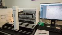
Multi-Mode Plate Reader / Imager (BioTek Cytation/BioStack)
- Plate reader for UV-Vis absorbance, fluorescence and luminescence detection
- Robotic microplate stacker
- Brightfield, phase, color brightfield, and fluorescence imaging of microplates, slides, and 35-mm dishes
- Objective lenses: 4x, 20x
Live-Cell Plate Imager / Incubator (BioTek Cytation/BioSpa)
- Long-term automated imaging of up to 8 plates for hours, days, or weeks
- CO2/humidity-controlled incubator for sample maintenance
- Robotic transfer of plates from incubator to temperature/CO2-controlled imaging platform
- Brightfield, phase, color brightfield, and fluorescence imaging of multi-well plates, slides, and 35-mm dishes
- Objective lenses: 4x, 10x, 20x, 40x, 60x
- Autoscratch device for automatic generation of scratch wounds in cell monolayers in 24- or 96-well plates
Accessories
- Specialized objective lenses for TIRF, intravital imaging, and other applications
- Cell culture room with laminar flow hood, cell culture incubator, water bath, and 4° refrigerator
- Lab bench with dissecting microscope, analytical balance, microcentrifuge, vortexer, water bath, and hotplate
- Stage-top incubators for temperature, humidity, and CO2 control during experiments
- Autoscratch (Agilent) monolayer scratch wound generator
- Microfluidics and perfusion equipment
- Syringe pump
- Peristaltic pump
- CellAsic ONIX microfluidic system for mammalian cells, yeast, or bacteria on specialized plates
- Animal procedure area with isoflurane anesthesia vaporizer, oxygen, warming pad, dissecting microscope
Image Analysis and Presentation
A Windows workstation with 256 GB RAM is available for image processing, analysis, and rendering. We provide consultation and training on analysis software. We also develop custom analysis workflows and scripts. Software includes:
Contact
Our Team
Acknowledgement and Co-authorship
We expect that the Confocal and Specialized Microscopy Shared Resource and its funding will be acknowledged in all publications that use our instruments, software, and/or expertise. This helps ensure our continued operation and acquisition of new instruments.
In addition, we welcome opportunities to provide significant intellectual and practical contributions that will lead to shared authorship.
How to cite us:
These studies used the Confocal and Specialized Microscopy Shared Resource of the Herbert Irving Comprehensive Cancer Center at Columbia University, funded in part through the NIH/NCI Cancer Center Support Grant P30CA013696.


