Neural Hijack: How Lung Cancer Rewires Itself to Spread to the Brain
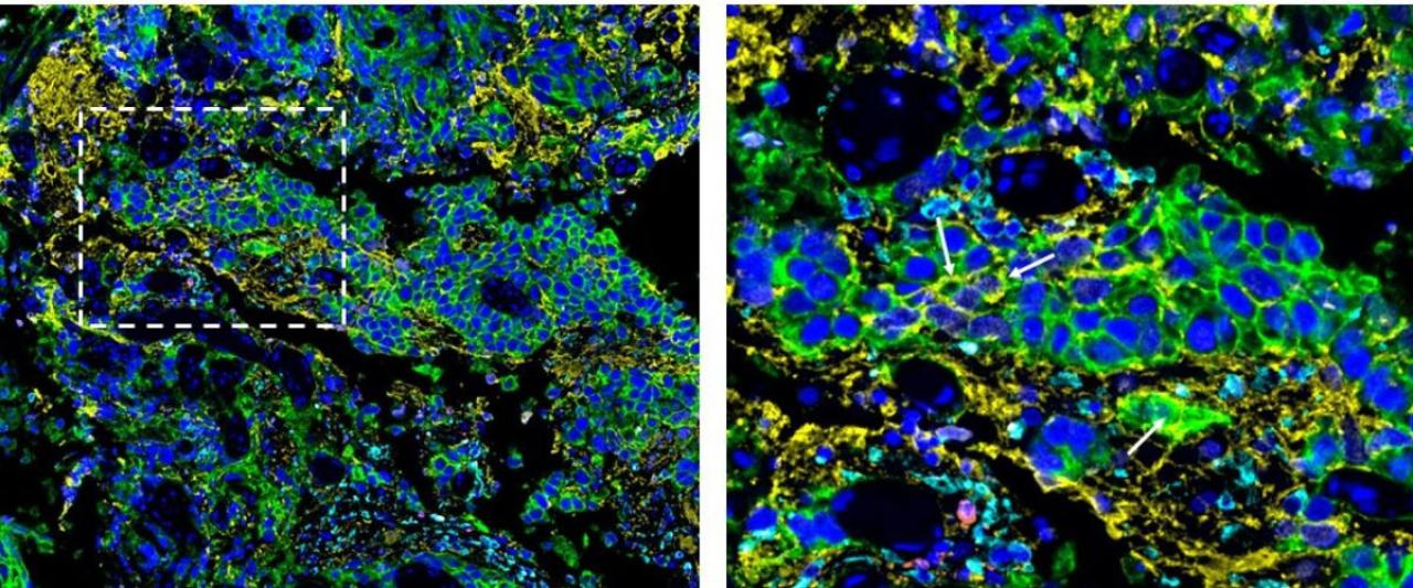
This image represents NSCLC metastatic site to the brain. When lung cancer cells (green) metastasize to brain, a small fraction of them adopts characteristics of glial cells (yellow), that form the major component of brain.

Combining cutting-edge single-cell and genomic profiling with innovative machine-learning approaches, researchers have identified genetic programs that drive one of the most challenging cancer complications: brain metastasis. The new work, published in Nature Medicine, points toward novel strategies to try to detect, prevent, and treat the spread of tumors to the brain.
Many tumors that start in other parts of the body can colonize the brain, but some do it much faster than others, including non-small cell lung cancer. "Up to 20% of patients with non-small cell lung cancer already have brain metastasis when they are diagnosed," says Benjamin Izar, MD, PhD, Milstein Family Aassociate Professor of Medicine at the Columbia University Vagelos College of Physicians and Surgeons and a member of the Herbert Irving Comprehensive Cancer Center (HICCC). "These metastases do not respond as well to treatment compared to areas outside the brain, which is a major unmet clinical need," adds Izar, senior author on the paper.
Novel sequencing techniques and machine learning enable analysis of millions of cells
Due to the lack of in-vivo models that accurately reflect human biology, Izar's lab turned towards characterizing the molecular landscapes of both primary lung tumors and brain metastases from a large library of patient samples at Columbia University Irving Medical Center (CUIMC). "We had access to extensive DNA sequencing data from over 12,000 patients who had non-small cell lung cancer, and this unprecedented dataset gives us confidence that our findings capture the true molecular signatures of brain metastases in this type of cancer," says Lindsay Caprio, an MD/PhD student in Izar's lab and one of the lead authors on the new paper. The scientists performed a series of tests on each sample, characterizing each individual cell in molecular detail.
The research was made possible by innovative new sequencing techniques pioneered in the Izar lab, which the team refined for the new project. "The ability to perform molecular profiling on individual cells at this level is enabled by a wave of technical innovations that we have been fortunate to contribute to in recent years," says Parin Shah, manager of the human immune monitoring core facility at the Columbia Center for Translational Immunology and a co-author on the study.
The massive sample size and exhaustive testing was only one part of the project. Analyzing the extensive dataset required breaking new ground in computational approaches as well. "A fundamental challenge in sequencing analysis is distinguishing cancer cells from other cell types," says Somnath Tagore, an associate research scientist in Izar's lab and a co-lead author on the study.
“To solve for this, we integrated multiple sequencing modalities, creating a novel machine learning tool that allowed us to more accurately differentiate cell types.”
Metastatic lung cancer cells behave like brain cells
Using this new advanced framework, the scientists made two major discoveries about the metastatic cells. First, brain metastases from non-small cell lung cancers showed extreme chromosomal instability, a phenomenon Izar's lab also saw previously in brain metastases from melanomas. Second, the results reveal a small group of cells in the original tumors that have gene expression patterns normally seen in neurons. "Lung cancers usually start in epithelial cells, but this rare group of cells seems to be behaving more like cells in the brain or nervous system," says Caprio. “This suggests that the tumor cells have undergone a profound shift in their transcriptional programs.” The team validated their findings with the help of CUIMC’s extensive pathology resources, as well as spatial data analysis in collaboration with Denis Schapiro.
This comprehensive analysis unravels the biology behind brain metastases in non-small cell lung cancer, giving researchers a clearer picture of how these metastases occur and behave – and potentially, how to stop them. Izar’s group is now spearheading the development of new targeted therapies aimed at preventing cancer cell migration to the brain. “We hope to not only deepen our understanding of metastases, but also open new avenues for intervention, and hope for patients,” says Izar.
References
Additional information
This paper, “Single-cell and spatial genomic landscape of non-small cell lung cancer brain metastases,” was published on February 27, 2025, in the journal Nature Medicine.
Funding
This study was supported by the National Institute of Health grants R37CA258829, R01CA266446, R01CA280414, U54CA274506, F30CA281104, T32AI148099, T32GM145440, P30CA013696, UL1TR001873, the Burroughs Wellcome Fund Career Award for Medical Scientists, a Velocity Fellows Award, the Louis V. Gerstner, Jr. Scholars Program, a Tara Miller Young Investigator Award by the Melanoma Research Alliance, a Tara Miller Team Science Award for Brain Metastasis Research by the Melanoma Research Alliance, a Pershing Square Sohn Cancer Research Alliance Award, the Cancer Research Institute (CRI) Lloyd J. Old STAR Program (CRI5579). This study was also supported by the Herbert Irving Comprehensive Cancer Center Human Tissue Immunology and Immunotherapy Initiative the Molecular Pathology Shared Resource and its Tissue Bank and the Human Immune Monitoring Core at Columbia University. Biospecimens and/or data used for this study were obtained from the Columbia University Biobank, which is partially supported by Columbia University’s Clinical and Translational Science Award.
